
Dissection activity
Cross-sectional multiplanar dissection
Fresh frozen adult human heads were dissected in the Center for Anatomy and Cell Biology, Division of Medical Sciences of the University of Brescia and Vienna.
The arterial system was injected via the common carotid and vertebral arteries with silicone rubber (Xiameter, Midland, MI, USA) stained with red Pintasol (Mixol Red E-L3mix, Kirchheim unter Teck, Germany).
At first, the project expected the section (on the axial, sagittal and coronal planes) of an entire cadaver head and neck with a dedicated band saw (Figure 1, A-C).
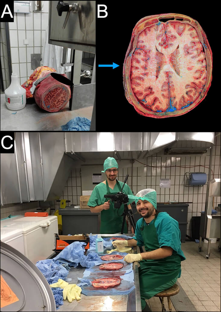
Figure 1, Cross-sectional multiplanar dissection. A: Band saw used at the Cadaver lab of the University of Vienna. B: Cross-sectional images on the axial plane. C: Preparation of anatomical specimens before photo shooting.
The following issues were highlighted during the first set of dissection:
- The twisting of the blade, due to pression and hardness of the bone cut, especially at the level of the temporal and sphenoid bones
- The suboptimal adequacy of the saw teeth for the purpose of cutting a human specimen, too little aggressive for the purpose of cutting a human specimen
- The excessive width of the cutting surface.
These issues led to the inadequacy of the cutting line, with not homogenous thickness of the slide (red square in Figure 2).
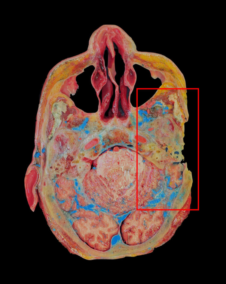
The cutting problems were overcome introducing a the PowerTek 350W saw with more aggressive teeth (cutting surface:1425X6,35 mm) (Figure 3 A-B).
A holder was specifically designed and built, with the aim of applying a constant pressure on the specimen while cutting (Figure 3A).
Furthermore, to ensure an easier cut, the volume of the specimen was reduced.
The final width of the slices was approximately 10 mm.
The slice positioned on a black surface and photos were taken with a CANON EOS 850d 18-55 f/4-5.6 IS and Manfrotto MKCOMPACTADV-BK photo tripod.

Figure 3, The PowerTek 350W band saw. A: the white arrow indicates the holder integrated on the band saw. B: cutting activity.
Endoscopic endonasal dissection
Fresh frozen adult human heads were dissected in the Center for Anatomy and Cell Biology, Division of Medical Sciences of the University of Brescia.
The arterial system was injected via the common carotid and vertebral arteries with silicone rubber (Xiameter, Midland, MI, USA) stained with red Pintasol (Mixol Red E-L3mix, Kirchheim unter Teck, Germany).
Dissections were performed using high-definition and 4K endoscopes and screen (Karl Storz®, Tuttlingen, Germany, and Olympus®, Tokyo, Japan, respectively), high-speed drill (Anspach®, High Wycombe, UK), and a complete set of endoscopic surgical instruments (Karl Storz®, Tüttlingen, Germany).
A step-by-step dissection of endoscopic endonasal approaches on the midline and coronal planes were performed. In particular:
- Functional endoscopic surgery including endoscopic opening of ethmoidal, frontal, maxillary (Figure 4 A), and sphenoid sinus
- Transfrontal approach
- Transcribriform approach (Figure 4 B-C)
- Transplanum and trascuberculum approach
- Transsellar approach
- Transdorsal approach
- Transclival approach
- Transodontoid approach
- Transmaxillary and trasnspterygoid approaches to the infratemporal fossa, ptarygopalatine fossa, cavernous sinus, meckel’s cave, and upper medial parapharyngeal space.
For all these approaches, images and videos were collected and edited.
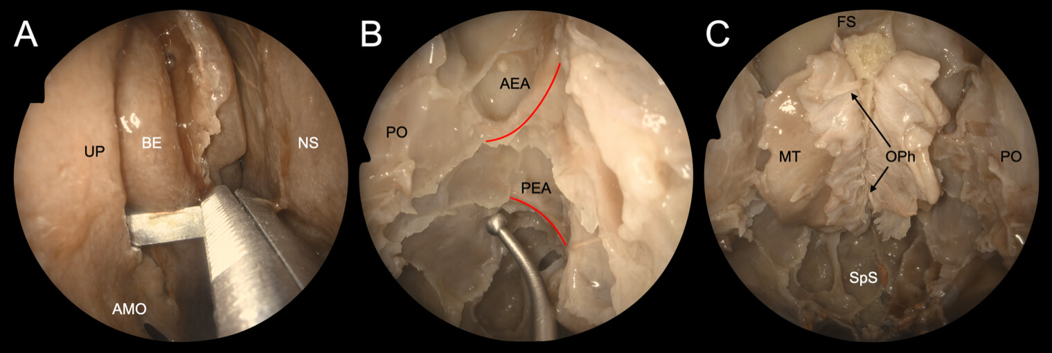
Figure 4, Images of endoscopic dissection. A: Right uncinectomy with a back-biting forceps (AOM – Accessory ostium of the maxillary sinus; BE – Bulla ethmoidalis; NS – Nasal septum; UP – Uncinate Process). B: Skull base exposure after ethmoidectomy (in red the AEA and PEA – Anterior and Posterior ethmoidal arteries; PO – Periorbit). C: Olfactory Phyla (OPh) and central anterior skull base (FS – Frontal Sinus; MT – Middle turbinate; PO – Periorbit; SpS – Spenoid sinus).
Open dissection
Fresh frozen adult human heads were dissected in the Center for Anatomy and Cell Biology, Division of Medical Sciences of the University of Brescia.
The arterial system was injected via the common carotid and vertebral arteries with silicone rubber (Xiameter, Midland, MI, USA) stained with red Pintasol (Mixol Red E-L3mix, Kirchheim unter Teck, Germany).
Dissections were performed using a complete set of open surgical instruments (Karl Storz®, Tüttlingen, Germany).
A step-by-step dissection of selected areas of the head and neck was performed, focusing on surgical details (Figure 5 B).
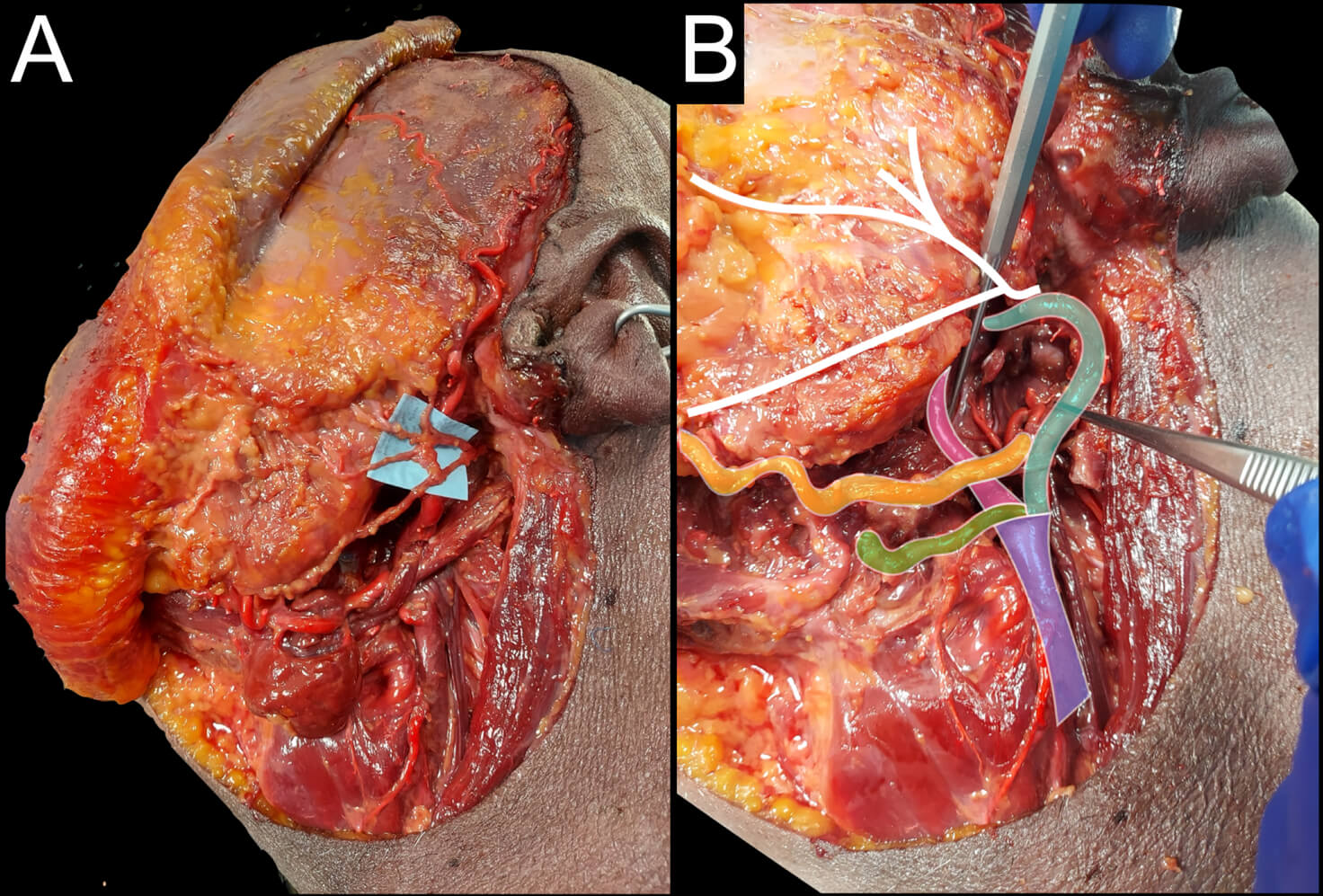
Figure 5, Open dissection images. A: dissection of the external tracts of the left facial nerve. B: Carotid network exposure after removal of the digastric muscle. In purple the common carotid artery, in green the lingual artery, in orange the facial artery, in blue the external carotid artery, and in pink the internal carotid artery. In white the facial nerve.
Three-dimensional models
Fresh-frozen adult human heads were dissected in the Laboratory of Endoscopic and Microsurgical Anatomy of the University of Brescia. The arterial system was injected via the common carotid and vertebral arteries with silicone rubber (Xiameter, Midland, MI, USA) stained with red Pintasol (Mixol Red E-L3mix, Kirchheim unter Teck, Germany). Dissections was performed using high-definition and 4K endoscopes and screen (Karl Storz®, Tuttlingen, Germany, and Olympus®, Tokyo, Japan, respectively), high-speed drill for endonasal surgery (Anspach®, High Wycombe, UK), and a complete set of endoscopic and open-field surgical instruments (Karl Storz®, Tüttlingen, Germany).
A dissection of the maxillary area was performed, with the aim of obtaining an anatomical piece composed by the maxillary bone, the ethmoid bone with the inferior and middle turbinate, and the pterygomaxillary junction, with the medial and lateral pterygoid laminae.
A tridimensional model of the surgical specimen was obtained with the structured light-based 3D Cronos Dual 2.0 Mpx scanner (Open Technologies 3D, Italy) (Figure 6 A).
The scanning field was set at 200 mm, with a working distance of 410 mm, and point spacing of 125 μm. The target was placed on a single plane 360-degrees rotating platform (Figure 6 B). The preliminary capture was sent to OpticalRevEng software (Open Technologies 3D, Italy) for the first 3D editing.
The 3D mesh obtained was uploaded in .obj format on an open-source system for processing and editing 3D triangular meshes (Meshlab, Visual Computing Lab) (Figure 6 C-D).
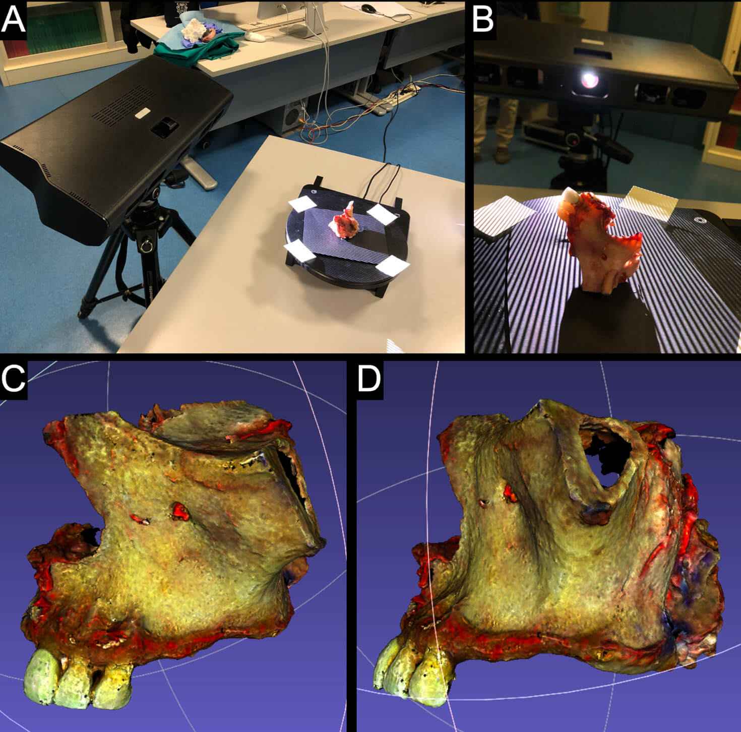
Figure 6, 3D scanning. A: scanning setting with the 3D Cronos Dual 2.0 Mpx scanner (Open Technologies 3D, Italy). B: 360-degree, single axis, rotating platform. C-D: anterolateral and posterolateral view of the 3D mesh obtained after maxillary bone scanning.
Structure of the website
Images derived from cross-sectional multiplanar dissection on the axial, sagittal and coronal plane have been organized in viewer boxes so that the user scroll them sequentially (Figure 7 A). Every image expects the presence of tags indicating the name of anatomical structure. Given the anatomical complexity of the head and neck area, anatomical structures of particular interest in the field of surgery have been favored for tagging (Figure 7 B).
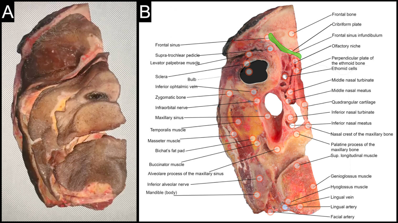
Figure 7, Sequential slices viewer. A: viewer box. B: Coronal image with tags indicating anatomical structures. In green, anterior skull base area linked to a multi-perspective panel (see figure 8).
Interactively, the user can explore areas of particular interest (Figure 7 B, green area) with panels that refer to the multi-perspective view of anatomical structures, combining endoscopic, open and radiological images (Figure 8 A-B).
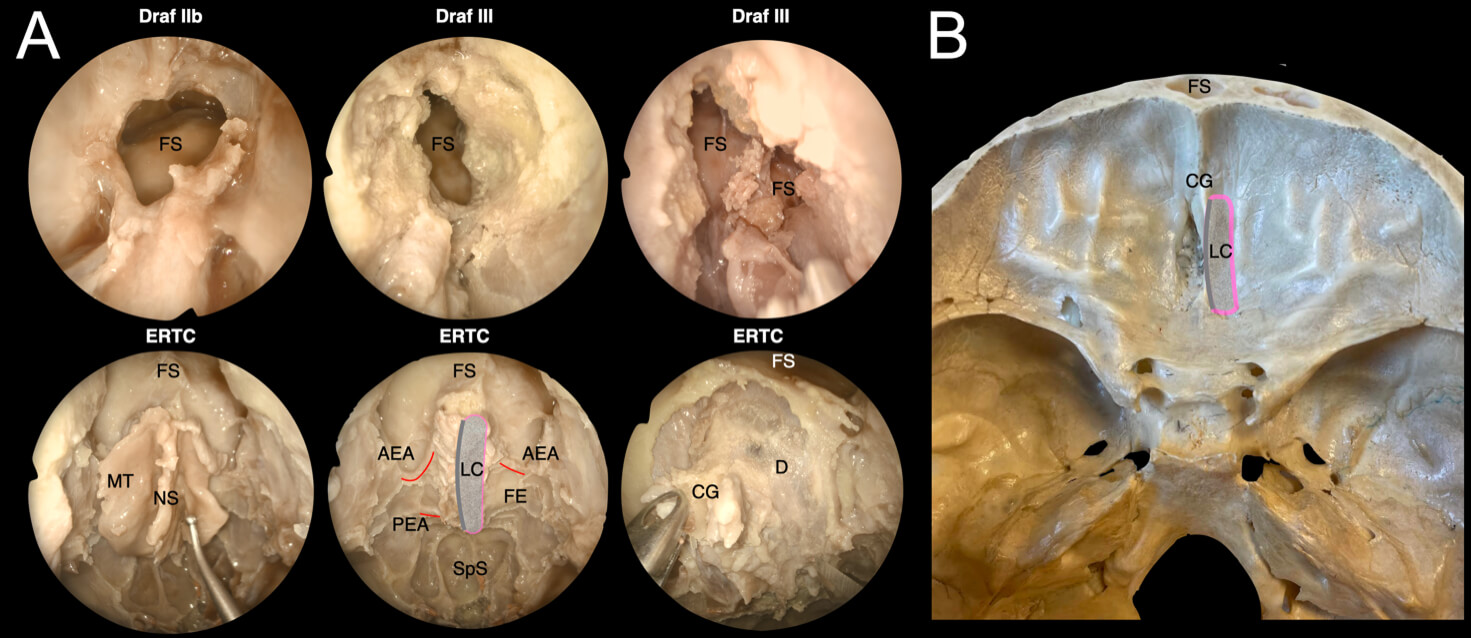
Figure 8, Multi-perspective panel. A: Series of step-by-step images of endoscopic dissection, including Draf IIb-III procedure and endoscopic resection with transnasal craniectomy (ERTC) (AEA – Anterior ethmoidal artery; CG – Crista galli; D – Dura mater; LC – lamina cribra, in grey; FE – Fovea ethmoidalis; FS – Frontal sinus; MT – Middle turbinate; NS – Nasal septum; PEA – Posterior ethmoidal artery; SpS – Spenoid sinus. B: Superior cranial view of the anterior skull base and frontal sinus.
Videos of dissection, surgical activity, and 3D models have been collected and uploaded on YouTube and then liked on the website.